> ## Documentation Index
> Fetch the complete documentation index at: https://docs.bioptimus.com/llms.txt
> Use this file to discover all available pages before exploring further.
# Tissue segmentation
> Separate tissue from background before feature extraction.
The tissue segmentation model identifies tissue regions in a slide, letting you discard background tiles before running [H-Optimus](/documentation/models/h-optimus) or [M-Optimus](/documentation/models/m-optimus). It is bundled with both model packages.
## Specifications
| Property | Value |
| -------------------- | -------------------------------------------------------------------- |
| Task | Binary tissue mask |
| Input | 512×512 RGB tile at 8.0 µm/px |
| Output | Flattened binary mask of length 262,144 (512×512), values 0.0 or 1.0 |
| SageMaker dispatch | `model_name: "tissue-seg"`, `mode: "prediction"` |
| Recommended instance | `ml.g5.xlarge` |
Run segmentation at the coarse 8.0 µm/px resolution first; it is cheap relative to per-tile feature extraction at 0.5 µm/px and can dramatically reduce the number of tiles you embed.
## Reshaping the output
The endpoint returns a flat array. Reshape it to a 512×512 mask:
```python theme={null}
import numpy as np
mask = np.array(result["output"]).reshape(512, 512)
```
## Deploy
Tissue segmentation has no separate deployment — it is **bundled with both the H-Optimus and M-Optimus packages** and served from the same endpoint. Deploy either model (see the [Deployment overview](/deployment/overview)) and `tissue-seg` is available alongside it.
## Using it in a pipeline
In practice you rarely call this endpoint directly — the Bioptimus SDK's tissue mask provider generates and caches masks for you, then filters tiles before feature extraction or prediction. See [Spatial transcriptomics](/guides/workflows/spatial-transcriptomics) and the [Bioptimus SDK](/guides/get-started/sdk).
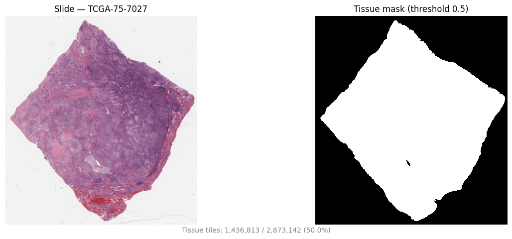
Once masks are generated, `bioptimus.utils.plot_slide_and_mask` renders the mask next to the slide thumbnail for a quick sanity check:
```python theme={null}
from bioptimus import utils
record = cohort[0]
utils.plot_slide_and_mask(record.wsi_path, record.mask_path, threshold=0.5)
```